When it comes to lung cancer, prevention is far and away the best medicine. But if this cancer does develop, an early, accurate and detailed diagnosis can help your doctor decide on the best approach for treatment.
If your doctor thinks you may have lung cancer, he or she will do several tests to find out for sure. If it is lung cancer, further testing can provide important information about what type of lung cancer it is and how far the disease has progressed.
According to the American Cancer Society (ACS) and the National Cancer Institute, tests used to diagnose lung cancer include:
Chest x-rays. This simple imaging test is often the first test a doctor recommends when lung cancer is suspected. If the x-ray image shows no unusual spots on the lungs, cancer is unlikely. If the image reveals an abnormality, the doctor may recommend more tests.
Computed tomography (CT) scans. These imaging tests rotate a narrow beam of x-rays around the body. The images are sent to a computer, which creates a detailed image of internal organs. The test can reveal the size, shape and location of a tumor. It can also reveal enlarged lymph nodes or growths in other organs, signs that cancer cells may be spreading beyond the original tumor.
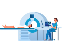
Magnetic resonance imaging (MRI) scans. MRIs produce more detailed images than CT scans, using powerful magnets, radio waves and computer processing. This test requires lying still inside a machine shaped like a tunnel, typically for 45 minutes to an hour. MRI scans are especially helpful for finding lung cancer that has spread to the brain or spinal cord.
Positron-emission tomography (PET) scans. PET scans are done with a special camera that picks up signals from radioactive substances. Before the test, a person is injected with a form of sugar with radioactive atoms. The camera then creates images that show where the radioactive substance is collecting. Cancer cells absorb much more of the sugar than normal cells, so areas with a heavy concentration of radioactive substance are likely to be cancer.
Bone scans can reveal unusual cell growth inside bones. This test may be recommended if other tests or symptoms suggest that lung cancer may have spread into the bones. For this test, a small amount of radioactive material is injected into a vein and a scanner outside the body measures radioactive buildup in the bones, a sign of possible cancer.
A closer look
If imaging tests show something that could be cancer, other tests may be recommended to confirm it. These more definitive tests include:
Sputum cytology. A sample of mucus coughed up from the lungs is examined under a microscope to look for cancer cells.
Needle biopsy. In this test, doctors take a tissue sample from the lungs using a tiny needle and examine it under a microscope for signs of cancer.
Mediastinoscopy. This test checks a tissue sample from the lymph nodes for cancer cells. The sample is taken through a small cut in the neck, where a hollow, lighted tube is inserted behind the chest bone.
Bronchoscopy. This test uses a flexible, lighted tube passed through the mouth into the airways of the lungs to look for tumors or blockages. The same instrument can be used to take tissue or fluid samples that can be checked for cancer cells.
Blood tests to check for signs that cancer may have spread to the bone marrow or liver.
Bone marrow biopsy to check a sample of bone marrow for cancer cells. The sample is usually taken from the pelvic bone with a long needle.
Endobronchial ultrasound or endoscopic esophageal ultrasound (EUS). These tests capture ultrasound images of the lungs and lymph nodes from inside the body, using a flexible, lighted tube fitted with ultrasound equipment. The tube is passed into the airways or the esophagus to get the imaging equipment as close as possible to the area being examined. These instruments can also be used to guide a needle to a lymph node to take a tissue sample that can be examined for signs that the cancer has spread beyond the lungs.
Thoracentesis or thoracoscopy to get information about the fluid that surrounds the lungs. A sample is drawn out with a needle and examined under a microscope.
Finding better ways
Many cases of lung cancer aren't found until they've been growing for some time, according to the ACS. This makes it more difficult to treat the cancer effectively.
When doctors find cancers early, they are often easier to treat. That's why the U.S. Preventive Services Task Force recommends that certain people at high risk of lung cancer be screened once a year for lung cancer. That includes people between ages 50 and 80 with a 20-year pack history who still smoke or who quit in the last 15 years. The test uses low-dose computed tomography.
Reviewed 3/27/2024
- American Lung Association. "Endobronchial Ultrasound (EBUS)." https://www.lung.org/lung-health-diseases/lung-procedures-and-tests/endobronchial-ultrasound-ebus.
- American Cancer Society. "MRI for Cancer." https://www.cancer.org/treatment/understanding-your-diagnosis/tests/mri-for-cancer.html.
- American Cancer Society. "Tests for Lung Cancer." https://www.cancer.org/cancer/small-cell-lung-cancer/detection-diagnosis-staging/how-diagnosed.html.
- American Cancer Society. "What's New in Lung Cancer Research?" https://www.cancer.org/cancer/lung-cancer/about/new-research.html.
- National Cancer Institute. "Bone marrow biopsy." https://www.cancer.gov/publications/dictionaries/cancer-terms/def/bone-marrow-biopsy.
- National Cancer Institute. "Small Cell Lung Cancer Treatment (PDQ)-Patient Version." https://www.cancer.gov/types/lung/patient/small-cell-lung-treatment-pdq.
- National Institute of Biomedical Imaging and Bioengineering. "Computed Tomography (CT)." https://www.nibib.nih.gov/science-education/science-topics/computed-tomography-ct.
- National Institute of Biomedical Imaging and Bioengineering. "Magnetic Resonance Imaging (MRI)." https://www.nibib.nih.gov/science-education/science-topics/magnetic-resonance-imaging-mri.
- U.S. Preventive Services Task Force. "Lung Cancer: Screening." https://www.uspreventiveservicestaskforce.org/Page/Document/UpdateSummaryFinal/lung-cancer-screening.